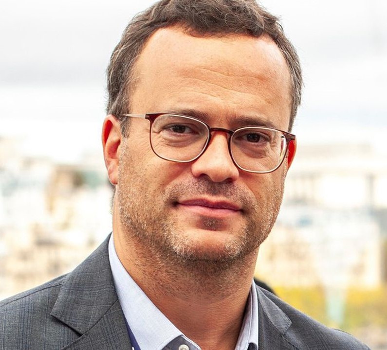
3 questions to Nor-eddine Regnard, Founder and Chief Medical Officer of Gleamer

Nor-eddine Regnard, co-founder and Chief Medical Officer at Gleamer, has been a radiologist in the Paris Hospitals for 4 years and teaches in prestigious faculties such as Pierre and Marie Curie University, Paris Descartes University, or the International University of Rabat in Morocco. In April 2018, he co-founded Gleamer alongside Christian Allouche (CEO) and Alexis Ducarouge (CTO), convinced that Artificial Intelligence can contribute to the resolution of complex medical problems.
Tell us more about your computer vision solutions. How does AI improve medical diagnosis in radiological examinations?
To understand this, you need to know the different steps in the work of a radiologist.
The radiologist starts by consulting the prescribing doctor's prescription to find out what is relevant to look for in order to answer the prescriber's question. His or her main job is to answer the question in the best way possible, which may be closed (e.g. suspicion of appendicitis?) or more open (e.g. abdominal pain).
He also has to look at all the images for other relevant abnormalities that are important to describe for the management of the patient or his follow-up. After the image acquisition phase, radiologists are asked to analyse the various images, usually at a high rate of speed, for abnormalities that are diagnostic or raise suspicion of diagnosis.
The radiologist can then compare these anomalies with previous examinations, measure their dimensions, and analyse the content and contours of the anomalies. This helps him to characterise the anomalies and arrive at a diagnosis.
Finally, after having observed everything and analysed the entire examination, the radiologist writes a detailed report.
Artificial Intelligence can help to improve the diagnosis of radiological examinations by
- by improving image quality, reducing radiation and saving time during image acquisition;
- by reliably, systematically and reproducibly detecting relevant abnormalities at any time of the day or night, anywhere in the world;
- by helping radiologists in the fine characterisation of anomalies;
- by carrying out automatic tasks such as segmentations, measurements, comparisons, which are often long and tedious for the radiologist;
- by helping to write a structured report.
Doctors and AI engineers work hand in hand at Gleamer. How does the combination of medical and AI expertise contribute to innovative and quality solutions?
At Gleamer, engineers and radiologists work together in several stages of the product life cycle:
- in the reflection on the relevant products in the radiologists' workflow;
- in the precise definition of the content of the products to have value and in the way they are built;
- in the definition of annotation instructions, in the quantitative and qualitative monitoring of these annotations;
- in the analysis of the software's performance as well as the clinical criteria to be favoured according to each product;
- in understanding user feedback;
- in the development and implementation of clinical studies, which are essential to obtain regulatory approval (CE, FDA) and to publicise the products;
- in the development of clinical cases;
- in the reflection on the design of products;
- in the reflection on data research.
This collaboration makes it possible to ensure that the products meet a real need for doctors, to monitor the medical quality of the software, to ensure its performance and to maximise the chances of the products being adopted by doctors.
What do you see as the most promising computer vision research for the medical sector?
Sectors where diagnosis is made partially or totally on images, such as radiology, nuclear medicine, ophthalmology, anatomopathology, dermatology, gastroenterology, etc.
Robotics, assisted by computer vision, is also a very promising field for surgery and interventional radiology.
In general, computer vision can also be used to identify prognostic factors for response to treatment on different types of images. This is particularly relevant in oncology and in the treatment of certain serious chronic diseases such as multiple sclerosis.
The US validation study paper for GLEAMER's BoneView AI solution entitled "Improving Radiographic Fracture Recognition Performance and Efficiency Using Artificial Intelligence" was awarded the Alexander Margulis Scientific Excellence Award for the best radiology paper of the year 2022 by the Radiological Society of North America (RSNA).
👉 Read more here.


